
ETINAL DETACHMENT - CAUSES AND HOW TO PREVENT IT (PART 1)
Retinal detachment describes an emergency situation in which a thin layer of tissue (the retina) at the back of the eye pulls away from its normal position. The vision process will be cut off and the patient’s vision will be partially or completely blurred depending on the degree of the retinal detachment This is a serious disease in ophthalmology with an emergency nature - if not treated in time, the patient may lose their vision permanently.
- What is retinal detachment?
Definition
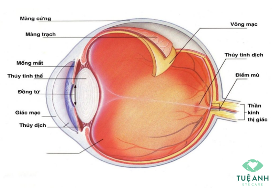
The retina is a thin layer of nerve tissue lining the inside of the eye, and is often compared to a film in a camera. This is because it has the function of receiving images and transmitting to the brain through the optic nerve.

When the retina detaches from its normal position, it causes retinal detachment, cutting off vision. You will experience partial or total blurred vision depending on the extent and of the detachment.
There are two types of retinal detachment:
- Primary retinal detachment, also known as retinal detachment with a tear, the only treatment is to perform surgery as soon as possible.
- Secondary retinal detachment: the main cause is inflammation (from uveitis) or elasticity (often seen in patients with diabetic retinopathy). The cause indicates the suitable treatment.
- Who is at risk of retinal detachment?
Factors that can cause this condition:
- Severe myopia with peripheral retinal degeneration (> -6.00D)
- Previously had retinal detachment in one eye
- Have had previous eye surgery (e.g. vitreous replacement surgery)
- Have diseases or disorders of the eye (such as retinal separation, uveitis, degenerative retina peripheral)
- Symptoms of primary retinal detachment
- Before retinal detachment occurs, the patient may see flies flying, spider web or flash.
- When retinal detachment is present, the patient will gradually lose vision, partially or completely depending on the degree and location of retinal detachment.
- The patient has absolutely no signs of pain or red eyes.
- In some cases, the condition appears suddenly which causes the patient to have rapid loss of vision. This is because the retina is teared, retinal blood vessels are damaged, causing bleeding into the vitreous chamber.
- Fundoscopy (which should only be performed by specialists): detects detachment and tear site.
- Eyeball ultrasound: helps to examine the retina and other intraocular structures, especially in cases of vitreous hemorrhage.

- How to recognize retinal detachment early
- See an ophthalmologist as soon as you notice symptoms of seeing a fly, or a flash, or a dark area in your vision.
- Take an annual eye exam, especially if you have higher risk for retinal detachment.
- Nearsighted patients should have bi-yearly eye exam
- When one eye already has retina detachment, it is necessary to check the other eye to detect early damage to the other retina - for laser preventive treatment.
- Wear goggles when playing sports and at working environments that may cause eye injury.
